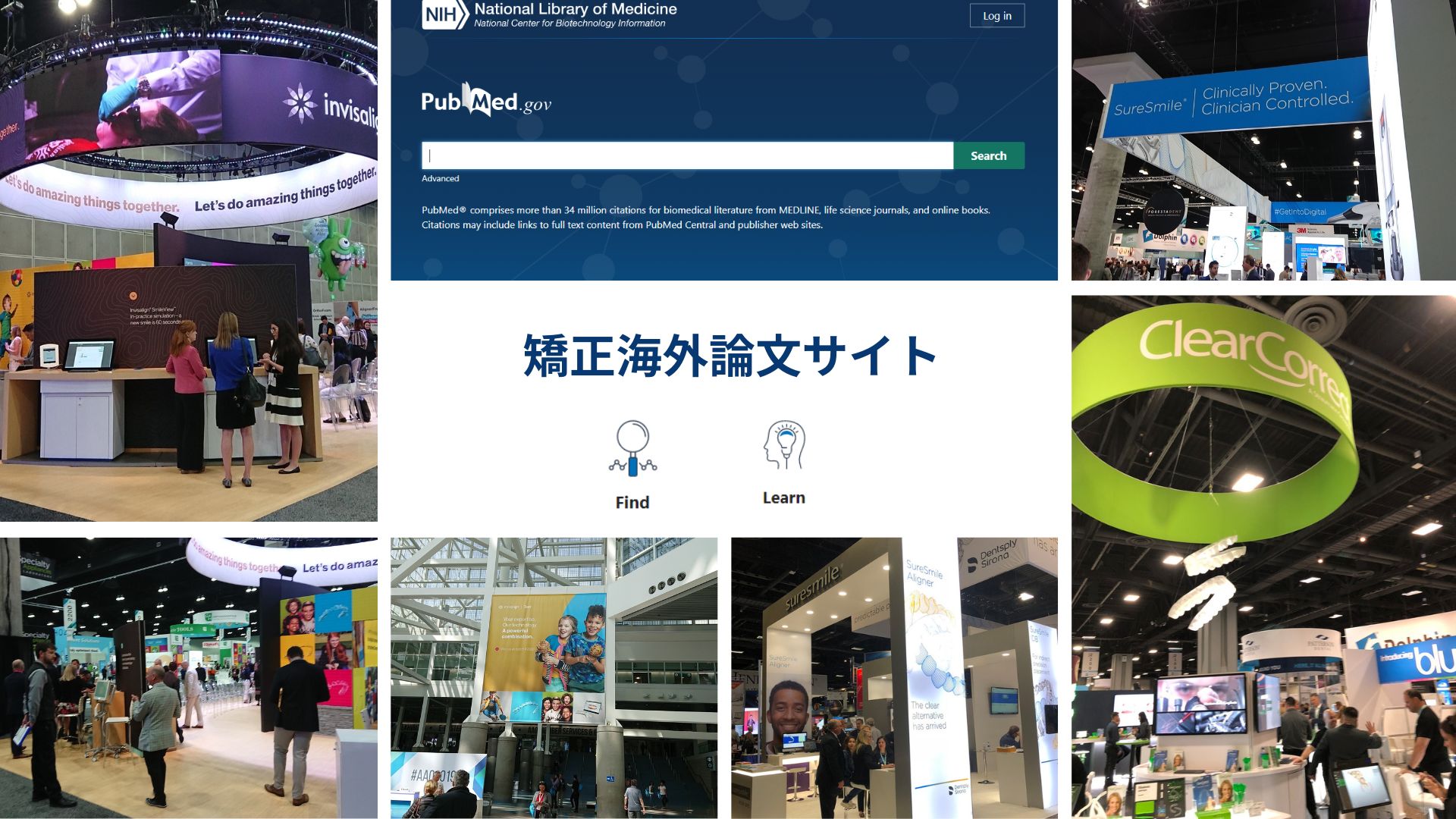
歯科医師、特に矯正医向けの最新矯正論文情報サイト
日常診療で多忙な歯列矯正医のためのドクター専用サイトです。
日々の診療や臨床、医学研究に役立つ最新海外論文をPubMedから情報を提供します。
アブストラクトだけでなく、結果の表やグラフ、診療前後の詳しい情報を日本語に訳して提供しています。
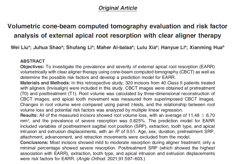
今回は、Angle Orthodontistに2021年に投稿された「Volumetric cone-beam computed tomography evaluation and risk factor analysis of external apical root resorption with clear aligner therapy.」をご紹介します。
お忙しい先生方の情報提供にお役立てください。
インビザライン歯根吸収の論文詳細
【タイトル】
Volumetric cone-beam computed tomography evaluation and risk factor
analysis of external apical root resorption with clear aligner therapy.
PMID:33826698 https://pubmed.ncbi.nlm.nih.gov/33826698/
【著者】Wei Liu; Juhua Shao; Shufang Li; Maher Al-balaa; Lulu Xia; Hanyue Li; Xianming Hua
【雑誌名】Angle Orthodontist, Vol 91, No 5, 2021
DOI: 10.2319/111820-943.1
インビザライン歯根吸収の要約
目的
クリアアライナ治療において、歯科用CTを用いて歯根吸収(EARR)の有病率と重症度を調査し、EARR発症の危険因子を特定しEARRの予測モデルを開発することである。
方法
クリアアライナー(インビザライン)治療の患者40名(クラスⅡ)の320本を対象と下後ろ向き研究である。
治療前(T0)と治療後(T1)に撮影した歯科用CT画像を用いた。
歯根の体積は歯科用CT画像を重ね合わせ計測した。
歯根体積の変化を対応のあるt検定で比較した。歯根体積の危険因子を特定するために重回帰分析を行った。
結果
測定したすべての歯で歯根体積の減少が見られた。
平均11.48±6.70㎣、重度の歯根吸収の有病率は0.625%であった。
年齢、性別、治療期間、治療前のSRPをモデルから除外した結果、EARRの発症は、治療後のSRP、抜歯、歯の種類、根尖の動きで説明された(R²=.51).
結語
アライナー治療におけるほとんどの切歯は軽度から中等度の歯根吸収を示し、重度の歯根吸収を示す割合はごくわずかであった。
治療後のSRP(最もEARRとの関連が強い )、抜歯、歯の種類、圧下と挺出はEARRの危険因子であることが示された。
インビザライン歯根吸収の論文を今すぐ読む!
引用文献
- Andreasen J. External root resorption: its implication in
dental traumatology. Paedodontics, periodontics, orthodontics
and endodontics. Int Endod J. 1985;18:109–118. - Weltman B, Vig KW, Fields HW, et al. Root resorptionassociated with orthodontic tooth movement: a systematicreview. Am J Orthod Dentofacial Orthop. 2010;137:462–476.
- Artun J, Van ’t Hullenaar R, Doppel D, et al. Identification oforthodontic patients at risk of severe apical root resorption.Am J Orthod Dentofacial Orthop. 2009;135:448–455.
- Puttaravuttiporn P, Wongsuwanlert M, Charoemratrote C, etal. Volumetric evaluation of root resorption on the upperincisors using cone beam computed tomography after 1 yearof orthodontic treatment in adult patients with marginal boneloss. Angle Orthod. 2018;88:710–718.
- Sharab LY, Morford LA, Dempsey J, et al. Genetic andtreatment-related risk factors associated with external apicalroot resorption (EARR) concurrent with orthodontia. OrthodCraniofac Res. 2015;18:71–82.
- Wierzbicki T, El-Bialy T, Aldaghreer S, et al. Analysis oforthodontically induced root resorption using micro-computedtomography (micro-CT). Angle Orthod. 2009;79:91–96.
- Tieu LD, Saltaji H, Normando D, et al. Radiologicallydetermined orthodontically induced external apical rootresorption in incisors after non-surgical orthodontic treatmentof Class II division 1 malocclusion: a systematic review. ProgOrthod. 2014;15:48.
- Sondeijker CFW, Lamberts AA, Beckmann SH, et al.Development of a clinical practice guideline for orthodonticallyinduced external apical root resorption. Eur J Orthod.2020;42:115–124.
- Walker SL, Tieu LD, Flores-Mir C. Radiographic comparisonof the extent of orthodontically induced external apical rootresorption in vital and root-filled teeth: a systematic review.Eur J Orthod. 2013;35:796–802.
- Dudic A, Giannopoulou C, Meda P, et al. Orthodonticallyinduced cervical root resorption in humans is associated withthe amount of tooth movement. Eur J Orthod. 2017;39:534–540.
- Elhaddaoui R, Qoraich HS, Bahije L, et al. Orthodonticaligners and root resorption: a systematic review. Int Orthod.2017;15:1–12.
- Li Y, Deng S, Mei L, et al. Prevalence and severity of apicalroot resorption during orthodontic treatment with clearaligners and fixed appliances: a cone beam computedtomography study. Prog Orthod. 2020;21:1.
- Krieger E, Drechsler T, Schmidtmann I, et al. Apical rootresorption during orthodontic treatment with aligners? Aretrospective radiometric study. Head Face Med. 2013;9:21.
- Aman C, Azevedo B, Bednar E, et al. Apical root resorptionduring orthodontic treatment with clear aligners: a retrospectivestudy using cone-beam computed tomography. Am JOrthod Dentofacial Orthop. 2018;153:842–851.
- Gay G, Ravera S, Castroflorio T, et al. Root resorptionduring orthodontic treatment with Invisalign: a radiometricstudy. Prog Orthod. 2017;18:12.
- Iglesias-Linares A, Sonnenberg B, Solano B, et al. Orthodonticallyinduced external apical root resorption in patientstreated with fixed appliances vs removable aligners. AngleOrthod. 2017;87:3–10.
- Ren H, Chen J, Deng F, et al. Comparison of cone-beamcomputed tomography and periapical radiography for detectingsimulated apical root resorption. Angle Orthod. 2013;83:189–195.
- Dudic A, Giannopoulou C, Martinez M, et al. Diagnosticaccuracy of digitized periapical radiographs validatedagainst micro-computed tomography scanning in evaluating orthodontically induced apical root resorption. Eur J Oral Sci.2008;116:467–472.
- Wang Y, He S, Yu L, et al. Accuracy of volumetric
measurement of teeth in vivo based on cone beam computer
tomography. Orthod Craniofac Res. 2011;14:206–212. - Maret D, Molinier F, Braga J, et al. Accuracy of 3D
reconstructions based on cone beam computed tomography.
J Dent Res. 2010;89:1465–1469. - Chien PC, Parks ET, Eraso F, et al. Comparison of reliability
in anatomical landmark identification using two-dimensional
digital cephalometrics and three-dimensional cone beam
computed tomography in vivo. Dentomaxillofac Radiol.
2009;38:262–273. - Koerich L, Weissheimer A, de Menezes LM, et al. Rapid 3D
mandibular superimposition for growing patients. Angle
Orthod. 2017;87:473–479. - Baumrind S, Korn EL, Boyd RL. Apical root resorption in
orthodontically treated adults. Am J Orthod Dentofacial
Orthop. 1996;110:311–320. - Aras I, Unal I, Huniler G, et al. Root resorption due to
orthodontic treatment using self-ligating and conventional
brackets: a cone-beam computed tomography study. J
Orofac Orthop. 2018;79:181–190. - Chang C, Liu Y. Root resorption during orthodontic treatment
with aligners. Chin J Orthod. 2018;25:191–195. - Pastro JDV, Nogueira ACA, Salvatore de Freitas KM, et al.
Factors associated to apical root resorption after orthodontic
treatment. Open Dent J. 2018;12:331–339. - Kato A, Ohno N. Construction of three-dimensional tooth
model by micro-computed tomography and application for
data sharing. Clin Oral Invest. 2009;13:43–46. - Durack C, Patel S, Davies J, et al. Diagnostic accuracy of
small volume cone beam computed tomography andintraoral periapical radiography for the detection of simulated external inflammatory root resorption. Int Endod J. 2011;44: 136–147. - Dudic A, Giannopoulou C, Leuzinger M, et al. Detection of
apical root resorption after orthodontic treatment by using
panoramic radiography and cone-beam computed tomography
of super-high resolution. Am J Orthod Dentofacial
Orthop. 2009;135:434–437. - Guo Y, He S, Gu T, et al. Genetic and clinical risk factors of
root resorption associated with orthodontic treatment. Am J
Orthod Dentofacial Orthop. 2016;150:283–289. - Deng Y, Sun Y, Xu T. Evaluation of root resorption after
comprehensive orthodontic treatment using cone beam
computed tomography (CBCT): a meta-analysis. BMC Oral
Health. 2018;18:116. - Lund H, Grondahl K, Hansen K, et al. Apical root resorption
during orthodontic treatment. A prospective study using
cone beam CT. Angle Orthod. 2012;82:480–487. - Castro IO, Alencar AHG, Valladares-Neto J, et al. Apical root
resorption due to orthodontic treatment detected by cone
beam computed tomography. Angle Orthod. 2013;83:196–
203. - Rudolph DJ, Willes MG, Sameshima GT. A finite element
model of apical force distribution from orthodontic tooth
movement. Angle Orthod. 2001;71:127–131. - Liu Y, Hu W. Force changes associated with different
intrusion strategies for deep-bite correction by clear aligners.
Angle Orthod. 2018;88:771–778. - Yamamoto T, Kinoshita Y, Tsuneishi M, et al. Estimation of
the remaining periodontal ligament from attachment-level
measurements. J Clin Periodontol. 2006;33:221–225.
記事監修


Dr. 堀井和宏(Kazuhiro Horii)
日本矯正歯科学会・臨床指導医(旧専門医)
American Association of Orthodontists
滋賀県大津市粟津町4-7
免責事項
本サイトは、PubMedに投稿されたオープンアクセスの査読論文をご紹介しております。
Discussionについては、すべての論文にagreeしているわけではありません。
しかし、「矯正海外論文サイト」は、最新矯正論文を提供するサイトとして矯正医の皆様がそれぞれ議論していただける一つのきっかけとなってくれたらと考えております。
また、内容についての疑問等は一切受け付けておりません。著者ご本人に記載の連絡先にお問い合わせをお願い致します。
さらに、学会発表や研修会においては本サイトの孫引きではなく必ず原著論文をご確認くださいますようお願い致します。
日本においてマウスピース型矯正はご存知の通り、完成薬機法対象外の矯正歯科装置であり、医薬品副作用被害救済措置制度の対象外となる場合がありますのでご注意ください。
矯正海外論文サイト公式インスタグラムのお知らせ
矯正海外論文サイトはInstagramを開設しております。
最新記事をお知らせする投稿がメインです。
最新論文のお知らせを受け取りたい方は 「矯正海外論文サイト」公式Instagramのフォローをぜひお願いします
なお、Instagramハイライトでは、
矯正治療の気になるKey Wordsを募集しております
矯正医の皆様のご回答をお待ちしております