歯科医師、特に矯正医向けの最新矯正論文情報サイト
日常診療で多忙な歯列矯正医のためのドクター専用サイトです。
日々の診療や臨床、医学研究に役立つ最新海外論文をPubMedから情報を提供します。
アブストラクトだけでなく、結果の表やグラフ、診療前後の詳しい情報を日本語に訳して提供しています。
今回は2022年にProgress in Orthodonticsに投稿された論文をご紹介します。
クリアアライナーで治療される叢生に対して、スペースを得るなどのために行われたIPRにおいて、クリンチェック上でプログラムされた量と実際に行われた量を歯牙ことに比較検討を行った研究です。
ん?と思った先生方、へえーと思った先生方いろいろだと思います。
論文紹介した後、この研究の方法論の問題など指摘しております。是非、最後までお読みください。
IPR論文
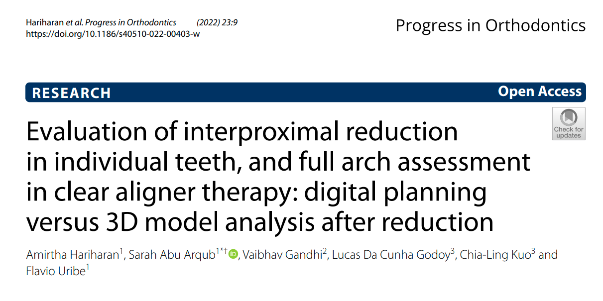
タイトル
Evaluation of interproximal reduction in individual teeth, and full arch assessment in clear aligner therapy: digital planning versus 3D model analysis after reduction.
PMID:35254555 https://pubmed.ncbi.nlm.nih.gov/35254555/
著者
Amirtha Hariharan1, Sarah Abu Arqub1*† , Vaibhav Gandhi2, Lucas Da Cunha Godoy3, Chia‑Ling Kuo3 and Flavio Uribe1
*Correspondence: sabuarqub@uchc.edu; super_orthodontist@hotmail.com
†Sarah Abu Arqub: Joint first author
1 Division of Orthodontics, Department of Craniofacial Sciences,
University of Connecticut Health, 263 Farmington Ave, Farmington, CT 06032, USA
雑誌名
Progress in Orthodontics (2022) 23:9
https://doi.org/10.1186/s40510-022-00403-w
論文要約
目的
臨床的に実施されたIPR(以下I-IPR)とクリンチェック上でプログラムされたIPR(以下P-IPR)との違いを比較検討すること。
材料と方法
75 名(男性 30 名、女性 45 名)、平均年齢 38±15 歳の被験者の治療前(T0)と治療後(T1)の ClinCheck® デジタルモデルを対象とした。
I-IPR 量を算出するため、Ortho Analyzer ソフトウェア (3Shape, Copenhagen, Denmark) を使用して,初期 (T0) と最終 (T1) STL モデルの上顎歯と下顎歯の第二小臼歯から対側第二小臼歯までの近遠心幅を測定した。
歯牙ごとに行われたI-IPRは、T0とT1における各歯牙の近遠心幅を比較することで求めた。
IPR の差 = (P-IPR)― (I-IPR).
さらにIPR論文を詳しく読む!
IPRとは
【IPRとは】
interproximal reductionの略語のことです。
・永久歯の隣接面の表面の縮小、解剖学的再形成、保護を含む補助的な臨床的処置[5]のことを指し、奥の隣接面の外側のエナメル質を約0.3~0.5mm削る方法である[6]。
【IPRのメリット】
・抜歯が望ましくない場合に、叢生を緩和し、歯の移動と整列を促進するためのスペースを得ることが出来る[7]。
・治療期間の短縮[6]、接触面積が増えるため安定性が増す[8]、ブラックトライアングルが減る[7]。
・Bolton Indexの不調和を軽減し[9]、歯周組織や歯科組織の健全性を損なわずに治療目的を達成するにに役立つ][10]。
引用文献
- Buschang PH, et al. Comparative time efciency of aligner therapy and
conventional edgewise braces. Angle Orthod. 2014;84(3):391–6. - Grünheid T, Loh C, Larson BE. How accurate is Invisalign in nonextrac‑
tion cases? Are predicted tooth positions achieved? Angle Orthod.
2017;87(6):809–15. - Lanteri V, et al. The efcacy of orthodontic treatments for anterior crowd‑
ing with Invisalign compared with fxed appliances using the Peer Assess‑
ment Rating Index. Quintessence Int. 2018;49(7):581. - Levrini L, Tieghi G, Bini V. Invisalign ClinCheck and the aesthetic digital
smile design protocol. J Clin Orthod JCO. 2015;49(8):518–24. - Peck H, Peck S. An index for assessing tooth shape deviations as applied
to the mandibular incisors. Am J Orthod. 1972;61(4):384–401. - Zachrisson BU, et al. Dental health assessed after interproximal enamel
reduction: caries risk in posterior teeth. Am J Orthod Dentofac Orthop.
2011;139(1):90–8. - Pindoria J, Fleming P, Sharma P. Inter-proximal enamel reduction in con‑
temporary orthodontics. Br Dent J. 2016;221(12):757–63. - Littlewood S, Kandasamy S, Huang G. Retention and relapse in clinical
practice. Aust Dent J. 2017;62:51–7 - Chee D, Ren C, Yang Y. An overview on interproximal enamel reduction.Dent Open J. 2014;1(1):14–8.
- Hellak A, et al. Infuence on interradicular bone volume of Invisalign treatment for adult crowding with interproximal enamel reduction: a retrospective three-dimensional cone-beam computed tomography study. BMC Oral Health. 2018;18(1):1–8.
- Zachrisson BU, Nyøygaard L, Mobarak K. Dental health assessed more than 10 years after interproximal enamel reduction of mandibular anterior teeth. Am J Orthod Dentofac Orthop. 2007;131(2):162–9.
- Radlanski RJ, et al. Plaque accumulations caused by interdental stripping. Am J Orthod Dentofac Orthop. 1988;94(5):416–20.
- Kravitz ND, et al. How well does Invisalign work? A prospective clinical study evaluating the efcacy of tooth movement with Invisalign. Am J Orthod Dentofac Orthop. 2009;135(1):27–35.
- De Felice ME, et al. Accuracy of interproximal enamel reduction during clear aligner treatment. Prog Orthod. 2020;21(1):1–7.
- Arman A, et al. Qualitative and quantitative evaluation of enamel after various stripping methods. Am J Orthod Dentofac Orthop, 2006;130(2): p.e7–131. e14.
- Kalemaj Z, Levrini L. Quantitative evaluation of implemented interproximal enamel reduction during aligner therapy: a prospective observational study. Angle Orthod. 2021;91(1):61–6.
- Laganà G, et al. Enamel interproximal reduction during treatment with clear aligners: digital planning versus OrthoCAD analysis. BMC Oral Health. 2021;21(1):1–6.
- Krieger E, et al. Invisalign® treatment in the anterior region. J Orofac Orthop/Fortschritte der Kieferorthopädie. 2012;73(5):365–76.
- Shigenobu N, et al. Patterns of dental crowding in the lower arch and contributing factors: a statistical study. Angle Orthod. 2007;77(2):303–10.
- Bishara SE, et al. Changes in the dental arches and dentition between 25 and 45 years of age. Angle Orthod. 1996;66(6):417–22.
- Tüfekçi E, et al. Opinions of American and Swedish orthodontists about the role of erupting third molars as a cause of dental crowding. Angle Orthod. 2009;79(6):1139–42.
- Danesh G, et al. Enamel surfaces following interproximal reduction with diferent methods. Angle Orthod. 2007;77(6):1004–10.
- Johner AM, et al. Quantitative comparison of 3 enamel-stripping devices in vitro: How precisely can we strip teeth? Am J Orthod Dentofac Orthop. 2013;143(4):S168–72.
- Hall NE, et al. Predictors of variation in mandibular incisor enamel thickness. J Am Dent Assoc. 2007;138(6):809–15.
- Macha ADC, et al.: Mesiodistal width and proximal enamel thickness of maxillary frst bicuspids. Braz Oral Res 2010;24:58–63.
- Woodsidea DG. The signifcance of late developmental crowding to early treatment planning for incisor crowding. Am J Orthod Dentofac Orthop. 2000;117(5):559–61.
- Gianelly AA. A strategy for nonextraction Class II treatment. In: Seminars in orthodontics. 1998;Elsevier.
- McGuinness NJ, et al. A stress analysis of the periodontal ligament under various orthodontic loadings. Eur J Orthod. 1991;13(3):231–42.
- Meeran NA. Cellular response within the periodontal ligament on appli‑ cation of orthodontic forces. J Indian Soc Periodontol. 2013;17(1):16.
- Boyd RL. Esthetic orthodontic treatment using the invisalign appliance for moderate to complex malocclusions. J Dent Educ. 2008;72(8):948–67.
- Hein C, Jost-Brinkmann P, Schillai G. The enamel surface quality after interproximal stripping—a scanning electron microscopic assessment of diferent polishing procedures. Fortschr Kieferorthop. 1990;51(6):327–35.
- Chudasama D, Sheridan JJ. Guidelines for contemporary air-rotor stripping. J Clin Orthod. 2007;41(6):315.
- Camardella LT, et al. Accuracy and reliability of measurements performed using two diferent software programs on digital models generated using laser and computed tomography plaster model scanners. Korean J Orthod. 2020;50(1):13–25.
- Murugesan A, Sivakumar A. Comparison of accuracy of mesiodistal tooth measurements made in conventional study models and digital models obtained from intraoral scan and desktop scan of study models. J Orthod. 2020;47(2):149–55
記事監修


最後にこのIPR研究の問題点の指摘
今回ご紹介した論文は、IPRの精度の評価は臨床的に非常に重要であるため、I-IPRとP-IPRとの違いを比較検証することを目的とした研究であった。
この研究は、矯正医によって異なるIPRの技法を用いていたため、潜在的な交絡因子が存在したことを考察で明らかにしている。また、IPRの技法によって精度が異なることが先行研究において明らかになっていると述べている。IPRの精度をI-IPRとP-IPRで比較し検証することが目的であるならば、IPRの技法は統制しなければならないと考えられる。
当院でのIPRにおいては本論文中に記載されている「モーター駆動の研磨ストリップ」を使用するとともに、クリンチェックの作成時に可及的に理想的な状態でIPRを行えるように各歯の移動のプランニングを行っている。また、IPRの実施時にIPRゲージを用いてP-IPRと一致するように行っている。したがって、I-IPRとP-IPRはほとんど変わらないと考えている。
IPRの技法を統一していないのであれば、用いたIPRの技法ごとにIPRの精度の評価をしなければならない。したがって、研究の方法論に問題があり、科学的な実証研究としてのエビデンスが欠けていると思われる。
主たる目的であるIPRのプロトコルについての言及はなされておらず、IPR量の算出方法や測定方法に重きを置いているという印象の研究であった。
さらに言うならば、考察においてIPRの精度について述べなくてはならないところ、より多くのP-IPRの処方についての解釈が述べられており、研究の目的とは異なる言及がなされていた。
免責事項
本サイトは、PubMedに投稿されたオープンアクセスの査読論文をご紹介しております。
Discussionについては、すべての論文にagreeしているわけではありません。
しかし、「矯正海外論文サイト」は、最新矯正論文を提供するサイトとして矯正医の皆様がそれぞれ議論していただける一つのきっかけとなってくれたらと考えております。
また、内容についての疑問等は一切受け付けておりません。著者ご本人に記載の連絡先にお問い合わせをお願い致します。
さらに、学会発表や研修会においては本サイトの孫引きではなく必ず原著論文をご確認くださいますようお願い致します。
日本においてマウスピース型矯正はご存知の通り、完成薬機法対象外の矯正歯科装置であり、医薬品副作用被害救済措置制度の対象外となる場合がありますのでご注意ください。
矯正海外論文サイト公式インスタグラムのお知らせ
矯正海外論文サイトはInstagramを開設しております。
最新記事をお知らせする投稿がメインです。
最新論文のお知らせを受け取りたい方は
「矯正海外論文サイト」公式Instagramのフォローをぜひお願いします
なお、Instagramハイライトでは、
矯正治療の気になるKey Wordsを募集しております
矯正医の皆様のご回答をお待ちしております